ANTI HER2 THERAPIES AGAINST BREAST CANCER

Overexpression of the HER2 protein occurs in approximately 25% of breast cancer cases, resulting in a therapeutic target of great interest against which various anti-HER2 therapies against breast cancer are being developed.

In this post we summarize the main types of anti HER2 therapies that are being incorporated into clinical practice in the approach to breast cancer.

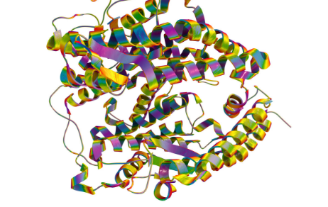
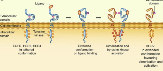
The HER2 protein, also known as ERBB-2, is a 185 kDa tyrosine kinase receptor composed of an extracellular ligand-binding domain, a transmembrane domain, and an intracellular domain with tyrosine kinase activity.

By binding to EGF (Epidermal Growth Factor) ligands, such as TGF-α , AR, EPR or NRG1 , among others, the formation of heterodimers is activated that activate various transduction cascades, resulting in increased proliferation, differentiation, cell mobility and survival.

Overexpression of this receptor and its constant activation leads to uncontrolled cell division and carcinogenesis.

After focusing on the HER2 receptor as a therapeutic target, various treatments aimed at blocking and deactivating it have been developed as a mechanism to treat HER2 + breast cancer.

There are currently three types of anti-HER2 therapies for breast cancer : monoclonal antibodies, TKIs (tyrosine kinase inhibitors) and ADCs (Antibody-drug conjugates).

MONOCLONAL ANTIBODIES (MAB)

Anti HER2 monoclonal antibodies are large molecules with monovalent affinity, produced using recombinant DNA technology.

Its mechanism of action is based on blocking the extracellular ligand binding domain of the HER2 receptor, thus reducing the neoplastic actions of HER2 heterodimers. At the same time, the HER2-antibody complex is internalized and destroyed by the target cell, reducing the number of available HER2 receptors.

Examples of monoclonal antibodies currently used as treatment in breast cancer include humanized antibodies trastuzumab and pertuzumab.

You can expand the information in this entry about Monoclonal Antibodies against breast cancer .

TYROSINE KINASE INHIBITORS (TKI)

Tyrosine kinase inhibitors are smaller molecules than monoclonal anti-HER2 antibodies, and their mechanism of action is based on the inhibition of phosphorylation of the intracellular tyrosine kinase domain of the HER2 receptor.

Most of these compounds target more than one HER receptor, allowing them to simultaneously block two or more components of the heterodimer.

The binding of these compounds to the HER2 receptor can be reversible as in the case of Lapatinib , or irreversible as in the case of Canertinib .

ANTIBODY-DRUG CONJUGATES (ADC)

ADCs are a special type of therapeutic antibody, consisting of an anti-HER2 monoclonal antibody conjugated to a chemotherapeutic drug. Its mechanism of action is based on the internalization of the antibody-drug complex and its intracellular cytotoxic activity.

The selection of the antigen for the development of this type of therapeutic agents is critical to achieve maximum specificity and consequently less toxicity, with optimal antigens being those that are expressed with high density on the surface of cancer cells, and do not do so in healthy cells.

An example of ADC against breast cancer would be trastuzumab emtansine (T-DM1).

5 METHODS TO QUANTIFY PROTEINS

The precise quantification of the proteins in a sample is essential for studying them in an infinity of research areas.

While the quantification of a specific protein can be carried out by means of tests such as Western Blot or ELISA , by mass spectrometry (MS) or by other methods such as those based on nanoparticles, there are tests that allow quantifying the concentration of total protein present in a shows.

This last case is the one we focus on this week, compiling a brief description of the 5 main methods to quantify total proteins .

1.- BICINCHONINIC ACID (BCA)
  • Concentration range : 20-2000 ug / ml
  • Features :
    • Developed in 1985
    • Colorimetric test where the absorbance will be proportional to the concentration of protein present in the sample
    • Two-step reaction:
      • Formation of protein-copper ion complexes
      • Formation of a Cu-BCA chelate resulting in an intense purple coloration that absorbs at 562nm
    • It is a recommended method in the case of samples containing> 5% detergents and / or denaturing agents such as urea or guanidinium chloride.
  • Limitations :
    • Samples containing substances that interact with copper, such as ammonia, like EDTA, reducing sugars, or lipids, may interfere with this assay.
2.- ULTRAVIOLET (UV) ABSORPTION
  • Concentration range : 0.1-100 ug / ml
  • Features :
    • This method, by measuring the characteristic absorption of tryptophan and tyrosine at 280nm, estimates the amount of protein present in the sample.
  • Limitations :
    • It is incompatible with protein extraction methods that use detergents and / or denaturing agents.
    • It is not a specific method for proteins, since other compounds that could be in the sample also absorb at 280nm (such as alcohols or nucleic acids, among others)
3.- BRADFORD OR COOMASIE BLUE TEST
  • Concentration range : 20-2000 ug / ml
  • Features :
    • Method described in 1976
    • It is characterized by being a quick, simple and compatible method with reducing agents used to stabilize proteins in solution.
    • The negatively charged Coomasie blue stain binds to positively charged proteins.
    • It mainly binds to arginine, tryptophan, tyrosine, histidine and phenylalanine residues.
    • The solution stain is red and absorbs at 465nm, while binding to the basic amino acids of a protein it turns blue and absorbs at 595nm.
    • The absorbance measurement is compared with the values ​​of a standard curve to determine the protein concentration of the sample.
  • Limitations :
    • Not valid for detecting proteins of <3kDa
    • It is incompatible with detergents such as SDS or Triton X-100
4.- LOWRY’S ESSAY
  • Concentration range : 10-1000 ug / ml
  • Features :
    • One of the most widely used methods to quantify proteins was developed in 1951
    • It is a very sensitive and precise test
    • Like the BCA assay, a two-step reaction occurs:
      • Formation of Cu-N complexes (present in the protein)
      • The tyrosine and tryptophan complexes react with the Folin-Ciocalteau rectifier, giving rise to a greenish-blue color that absorbs between 650 and 750nm.
    • The absorbance measurement is compared with the values ​​of a standard curve to determine the protein concentration of the sample.
  • Limitations :
    • It is incompatible with certain chemical reagents in common use such as Tris, EDTA, DDT, 2-mercaptoethanol, carbohydrates, etc.
5.- CBQCA (3- (4-CARBOXYBENZOYL) QUINOLINE-2-CARBOXYDEHYDE) ASSAY
  • Concentration range : 100 ng – 1500 ug / ml
  • Features :
    • It is a highly sensitive fluorogenic agent that is used for the detection of primary amines in proteins.
    • The intensity of the fluorescence emitted will be directly proportional to the concentration of protein present in the sample.
    • Does not interact with lipid compounds
  • Limitations :
    • The result depends on the number of certain amino acids present in the protein.