Stages of Meiosis

Meiosis describes the process of cell division by which gametes are formed. In this process, we start with a cell with twice the normal amount of DNA and end up with 4 non-identical haploid daughter gametes after two divisions. There are six stages of Meiosis within each of the divisions, namely prophase, prometaphase, metaphase, anaphase, telophase, and cytokinesis. In this article, we will look at the stages of meiosis and consider their importance in disease.

Meiosis I

In meiosis I, the homologous chromosomes separate in two cells, so that there is one chromosome (consisting of two chromatids) per pair of chromosomes in each daughter cell, that is, two chromosomes in all.

Prophase I

Before prophase, the chromosomes replicate to form sister chromatids. Initially, there are four chromatids (c) and two chromosomes (n) for each of the 23 pairs of chromosomes (4c, 2n). The nuclear envelope disintegrates and the chromosomes begin to condense. Spindle fibres appear that are important for the successful division of chromosomes.

To further increase genetic diversity, homologous chromosomes swap small parts of themselves, so that one chromosome contains both maternal and paternal DNA. This process is known as crossing over, and the points where this occurs on a chromosome are called chiasmata.

Prometaphase I

Spindle fibres attach to chromosomes at points along the chromosomes called centromeres. As this happens, the chromosomes continue to condense.

Metaphase I

Maternal and paternal versions of the same chromosome (homologous chromosomes) line up along the equator of the cell. A process called independent assortment occurs: This is when the maternal and paternal chromosomes line up randomly on either side of the equator. This in turn determines which gamete chromosomes are assigned to, leading to genetic diversity among offspring.

Anaphase I

Here, each of the homologous chromosomes is drawn to opposite poles of the cell as the spindle fibres retract. This equally divides the DNA between the two cells that will form.

Telophase I and Cytokinesis I

During telophase, I, the nuclear envelope reforms and the spindle fibres disappear. In cytokinesis I, the cytoplasm and the cell divide result in two cells that are technically haploid: there is one chromosome and two chromatids for each chromosome (2c, n).

Meiosis II

Prophase II and Prometaphase II

These stages are identical to their counterparts in meiosis I.

Metaphase II

In metaphase II, the chromosomes line up in a single file along the equator of the cell. This is in contrast to metaphase I, where the chromosomes line up in homologous pairs.

Anaphase II

Sister chromatids are then attracted to opposite poles of the equator.

Telophase II

This stage is the same as telophase I.

Cytokinesis II

Again, the cytoplasm and the cell divide produce 2 non-identical haploid daughter cells. Since this happens in both cells produced by meiosis I, the net product is 4 non-identical haploid daughter cells, each containing a chromosome consisting of one chromatid (1c, 1n). These are fully formed gametes.

Spliceosome

Abstract

The spliceosome is a large RNA-protein complex that catalyzes the removal of introns from nuclear pre-mRNA. A wide range of biochemical and genetic studies show that the spliceosome comprises three major RNA protein subunits, the small nuclear ribonucleoprotein (snRNP) particles U1, U2, and [U4/U6.U5], and an additional group of splicing proteins. not snRNP. factors Rapid progress is being made in unravelling the interactions that take place between these factors during the splicing reaction.

The emerging picture of the spliceosome reveals a highly dynamic structure that assembles into pre-mRNA transcripts in a stepwise pathway and is organized, at least in part, by complex RNA base-pairing interactions between small nuclear RNAs (snRNAs) and the intron substrate. Many of these interactions can be detected in both mammalian and yeast spliceosomes, suggesting that the basic splicing mechanism is ancient and largely conserved during evolution.

What are spliceosomes?

Spliceosomes are huge multimegadalton ribonucleoprotein (RNP) complexes found in eukaryotic nuclei. They assemble into RNA polymerase II transcripts from which they extract RNA sequences called introns and splice flanking sequences called exons. This so-called pre-messenger RNA (pre-mRNA) splicing is an essential step in eukaryotic mRNA synthesis. Each human cell contains approximately 100,000 spliceosomes, which are responsible for removing more than 200,000 different intron sequences. Human cells contain two types of spliceosomes: the major spliceosome, which is responsible for removing 99.5% of introns, and the minor spliceosome, which removes the remaining 0.5%.

How did the various parts of the spliceosome get their names?

U snRNAs were originally discovered as abundant small uridine-rich RNA molecules present in mammalian nuclei and were initially numbered in order of their apparent abundance. U1, U2, U4, U5, U6, U11, and U12 were later found to be components of the splicesome. The U7 snRNA is required for processing the 3′ end of histone mRNA; the other abundant U snRNAs (U3, U8, U9, and U10) are all involved in ribosome biogenesis. U4atac and U6atac are much less abundant than other spliceosomal snRNAs, so they were only discovered and named when it was realized that there must be other snRNAs that recognize the minor intron class.

The first and last two DNA nucleotides of minor introns are usually AT and AC, respectively, hence the names U4atac and U6atac. Many spliceosomal proteins have PRP names, e.g. Prp2, Prp5, Prp8, etc. In yeast, mutations in these genes lead to “mRNA pre-processing” defects. Confusingly, orthologous genes may have different PRP names in Saccharomyces cerevisiae and Schizosaccharomyces pombe because the original mutational screens were done around the same time and a unified naming system has not yet been devised.

Other core splicing proteins include CWC (complexed with CDC5), CWF (complexed with CDC five), SPF (Pichia farinosa killer toxin sensitivity), SYF (synthetic lethal with cdcforty). Complex nineteen (NTC) is a large protein-only subcomplex named for its most abundant component, Prp19, while another small protein-only complex known as NTR (related to complex nineteen) contains factors involved in spliceosome disassembly. Some of the major spliceosomal proteins were first discovered in invertebrates.

The seven Sm proteins, which form a ring surrounding a specific binding site on almost all spliceosomal snRNAs, were named after the patient (Smith) with whose autoimmune antibodies they react. A similar set of proteins (Lsm, for “Sm-like”) was later found to surround the U6 and U6atac snRNAs, the only two spliceosomal snRNAs that lacked a consensus Sm binding site. Two additional large classes of metazoan splicing factors are the hnRNP proteins, so named because they are found associated with heterogeneous nuclear RNA (hnRNA), and the SR proteins, named for a carboxy-terminal domain rich in arginine-serine dipeptides ( RS).

How does the spliceosome do its job?

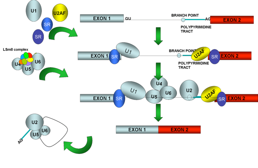
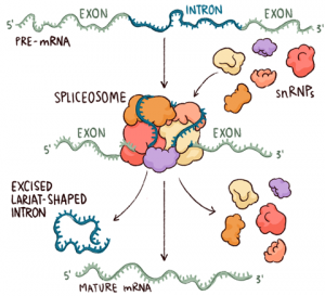
Spliceosomes must remove non-coding introns from precursor transcripts and rejoin flanking exons to create mature spliced ​​mRNAs. To do so, splicing machinery assembles step by step at the ends of introns, with U1 snRNP recognizing the start of an intron (5′ splice site, the donor site) and U2 snRNP recognizing a feature (the donor site). branch) at the other end in the vicinity of the 3′ splice site (acceptor site).

After numerous structural rearrangements involving both the addition of new components and the expulsion of many others, splicing occurs in two chemical steps: first, cleavage at the 5′ splice site along with the formation of a lariat structure. in which the first nucleotide of the intron is linked via a 2′–5′ phosphodiester bond to the adenosine branch site; and second, ligation of the two exons, along with cleavage at the 3′ splice site. The spliceosome then disassembles from the excised intron, which subsequently debranches and degrades.

How do spliceosomes affect gene expression?

Because the vast majority of protein-coding genes in humans contain introns (usually 9 or 10, but some have more than 100!), splicing is an essential step in gene expression. High-throughput sequencing has now revealed that ~95% of human genes are also subject to alternative splicing, allowing the synthesis of many different mRNAs from a single DNA gene. By encoding alternative protein isoforms or harbouring different regulatory sequences in their untranslated regions, alternatively spliced ​​mRNAs greatly enhance biological complexity.

The act of splicing itself also has important consequences for gene expression beyond intron removal. By stably depositing proteins that accompany mRNPs in the cytoplasm (e.g., the exon-joining complex, EJC) into exons, splicing can affect subcellular localization, translational efficiency, and decay kinetics of mRNP. mRNA. In particular, mRNA decay driven by the location of EJC relative to the stop codon is a crucial mediator of cellular protein abundance.

Are spliceosomes associated with any disease?

Many human diseases are caused by the misplacing of a single gene or by the dysregulation of the entire spliceosome. About 35% of human genetic disorders are caused by a mutation that disrupts the splicing of a single gene. Such mutations can add/delete a single splice site (eg, α or β thalassemia) or change the balance of alternative splicing by affecting the inclusion/exclusion of a cassette exon (eg, frontotemporal dementia driven by a misplacing of tau). Some misplacing events generate an isoform of mRNA that is subject to rapid degradation.

Single point mutations affecting splicing can result in large changes in both protein structure and protein abundance. Other diseases are caused by mutations in the splicing proteins themselves, affecting the splicing of many transcripts. For example, mutations in several core splicing proteins (eg, Prp8, Prp3, Prp31, and Brr2) have been shown to cause autosomal dominant retinitis pigmentosa. Mutations in splicing factor 3B subunit 1 (SF3B1) and U2 helper factor 35 (U2AF35) are frequently associated with chronic lymphocytic leukaemia and myelodysplasia. Other types of cancer are associated with dysregulation of splicing factor levels. Therefore, the spliceosome has recently emerged as a new target for the development of new cancer therapies.

What is left to explore?

Due to its highly dynamic and complex nature, an atomic-level structure of the spliceosome remains an elusive goal. However, much progress has recently been made by crystallizing subsets of spliceosomal components, including the U1 and U4 snRNPs and the central core protein Prp8. Other important questions concern the exact molecular mechanisms by which spliceosomes achieve high splicing precision while allowing flexibility in splice site choice to enable alternative splicing. To answer these questions, new tools such as single-molecule microscopy, bioinformatics, and high-throughput methods for determining protein-protein, protein-RNA, and RNA-RNA interaction dynamics are increasingly being developed and applied.

Spermatogenesis

Introduction

Spermatogenesis, is the origin and development of sperm within the male reproductive organs, the testes. The testes are composed of numerous thin, tightly coiled tubules known as the seminiferous tubules; sperm are produced within the walls of the tubules. Within the walls of the tubules, too, there are many randomly scattered cells, called Sertoli cells, that function to support and nourish immature sperm by providing them with nutrients and blood products.

As the young germ cells grow, Sertoli cells help transport them from the outer surface of the seminiferous tubule to the central canal of the tubule. The testes continually produce sperm, but not all areas of the seminiferous tubules produce sperm at the same time. An immature germ cell takes up to 74 days to reach final maturation and during this growth process, there are intermittent resting phases.

The immature cells (called spermatogonia) are all derived from cells called stem cells in the outer wall of the seminiferous tubules. Stem cells are composed almost entirely of nuclear material. (The nucleus of the cell is the portion that contains the chromosomes.) Stem cells begin their process by multiplying in the cell duplication process known as mitosis. Half of the new cells in this initial culture become future sperm cells and the other half remain as stem cells, so there is a constant supply of additional germ cells.

Spermatogonia destined to become mature sperm are known as primary sperm. These move from the outer portion of the seminiferous tubule to a more central location and coalesce around the Sertoli cells. The primary sperm cells then develop somewhat by increasing the amount of cytoplasm (substances outside the nucleus) and structures called organelles within the cytoplasm.

After a resting phase, the primary cells divide into a form called secondary sperm. During this cell division, a breakdown of nuclear material occurs. In the nucleus of primary sperm, there are 46 chromosomes; in each of the secondary spermatozoa, there are only 23 chromosomes, as there are in the egg. When the egg and sperm combine and their chromosomes unite, the characteristics of both individuals mix and the new organism begins to grow.

The secondary sperm must still mature before it can fertilize an egg; maturation involves certain changes in the shape and form of the sperm cell. The nuclear material becomes more condensed and oval in shape; this area develops as the sperm head. The head is partially covered by a cap, called an acrosome, which is important in helping sperm enter the egg. Attached to the opposite end of the head is the tailpiece.

The tail is derived from the cytoplasm of the secondary sperm. In mature sperm, it consists of a long, thin bundle of filaments that propel the sperm by their undulating motion. Once the sperm have matured, they are transported through the long seminiferous tubules and stored in the epididymis of the testicles until they are ready to leave the male body.

Characteristics of normal spermatogenesis based on histological sections

– Diameter of the seminiferous tubule 180 μm minimum

– Presence of spermatogonia type A pale, type A dark, type B

– Presence of primary and secondary spermatocytes

– Differentiation of spermatids

– Spermiation zones

– Score count of at least 8 (see section – “Score count for the evaluation of spermatogenesis”).

– lumen of seminiferous tubule

– Normal distribution of lipids in the cytoplasm of Sertoli cells

– Presence of spermatogenesis stages

– Formation of germ cell clones

– Thickness of the lamina propria of the seminiferous tubule of 8 μm or less

– Structure and normal distribution of Leydig cells

Kinetics of spermatogenesis

Spermatogenesis begins during puberty and continues throughout life and into old age due to the inexhaustible reservoir of stem cells. A large number of germ cells develop and are released from the seminiferous tubules. The process of spermatogenesis is highly organized: the spermatogonia divide continuously, in part spermatogonia remain and in part give rise to spermatogenesis. Originating from the division of the spermatogonia, the groups of cells migrate from the basal to the adluminal position of the germinal epithelium.

Differently developed cell groups are found in a section of a seminiferous tubule and contribute to the typical appearance of the germinal epithelium. Six of these typical aspects were described in the human testis as “stages of spermatogenesis”. In any given region of the germinal epithelium, the same typical appearances of groups of germ cells appear every 16 days. This period of time is called the “seminiferous epithelial cycle”.

The development of a type A spermatogonium to mature spermatids requires 4.6 cycles, e.g. 74 days Mature spermatids released from the germinal epithelium as spermatozoa are transported through the epididymal duct system for an additional 12 days. Therefore, a minimum of 86 days should be calculated for a complete spermatogenetic cycle from spermatogonium to mature sperm.

Alterations of spermatogenesis

The proliferation and differentiation of male germ cells and the intratesticular and extratesticular mechanisms of regulation of spermatogenesis can be altered at all levels. This may occur as a result of environmental influences or may be due to diseases that directly or indirectly affect spermatogenesis. In addition, different nutritional and therapeutic substances, drugs, hormones and their metabolites, different toxic substances or X-rays can reduce or destroy spermatogenesis. Finally, also a fairly simple nose since the increase in temperature reduces the spermatogenetic activity of the testicles.

Under these negative influences, testicles respond relatively monotonically by reducing spermatogenesis. This can be expressed in the reduced number of mature spermatids, in spermatid malformation, lack of spermiation, impaired meiosis, arrest of spermatogenesis at the primary spermatocyte stage, reduced multiplication or apoptosis of spermatogonia. If the spermatogonia survive, then spermatogenesis can be rescued.

Otherwise, spermatogenesis ceases, and the shadows of the seminiferous tubules remain. Alterations in spermatogenesis are evaluated in histological sections of testicular biopsies. The most suitable technique is the semi-thin cut of the material embedded in epoxy resin. In a semi-thin section, all the details of the testicle cells can be optimally evaluated due to their excellent preservation.